Structurated Scintillator
04.11.2019, 14:47
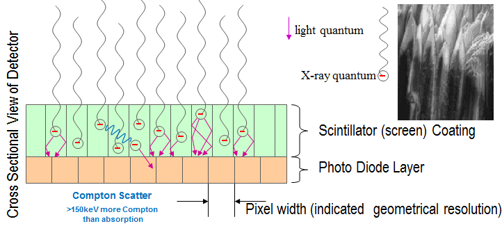
A way to overcome the influence of the 360° distrubuted light quants is to guide the light inside structure, e.g. chanels where the light quants are reflected at the walls.
The classical approach is CsI (Caesium iodide), doped with sodium (CsI (Na)), or doped with thallium (CsI(Tl)) for higher light output in the green region (550nm). CsI can be grown as crystals and then have a needle structure which forces the light inside the stucture.

Due to the better efficiency CsI is taken often in medical applications. But there are two issues with CsI with higher energies (>60keV):
a) CsI tends to get Burn In with creates ghosting (sometime you can still see the object from last session)
b) Compon Scatter is the dominating effect at energies above 150keV and the needle structure is obsolete
Here is a strange example for Burn In with a brand new detector with CsI. A 6mm steel plate with a on-top 5mm steel plate with test-welds and a Wire IQI is exposed with 160kV at 3mA for 300s. The contrast from the plate with the welds to the base plate is 12,500 grey values (from 65,535).
Then X-ray is shut down and 180s later this image is taken (0 kV, 0mA):
You can still see all what was in the image before (and even some more), but to be fair: The contrast is 43 grey values only.
Now all objects are removed out of the beam.
After further 120s X-Ray is turned on with same dose as above and now you can see the objects from before with a good contrast (620 grey values):
There is still 5% of contrast from the image before ; if you have to meet 2-2T you have to detect flaws with 2% contrast
  .
.
The classical approach is CsI (Caesium iodide), doped with sodium (CsI (Na)), or doped with thallium (CsI(Tl)) for higher light output in the green region (550nm). CsI can be grown as crystals and then have a needle structure which forces the light inside the stucture.

Due to the better efficiency CsI is taken often in medical applications. But there are two issues with CsI with higher energies (>60keV):
a) CsI tends to get Burn In with creates ghosting (sometime you can still see the object from last session)
b) Compon Scatter is the dominating effect at energies above 150keV and the needle structure is obsolete
Here is a strange example for Burn In with a brand new detector with CsI. A 6mm steel plate with a on-top 5mm steel plate with test-welds and a Wire IQI is exposed with 160kV at 3mA for 300s. The contrast from the plate with the welds to the base plate is 12,500 grey values (from 65,535).
Then X-ray is shut down and 180s later this image is taken (0 kV, 0mA):
You can still see all what was in the image before (and even some more), but to be fair: The contrast is 43 grey values only.
Now all objects are removed out of the beam.
After further 120s X-Ray is turned on with same dose as above and now you can see the objects from before with a good contrast (620 grey values):
There is still 5% of contrast from the image before
Last edited by Klaus on 30.08.2023, 17:05, edited 2 times in total.
Reason: Repaired links to the images
Reason: Repaired links to the images
Re: Structurated Scintillator
04.11.2019, 14:59
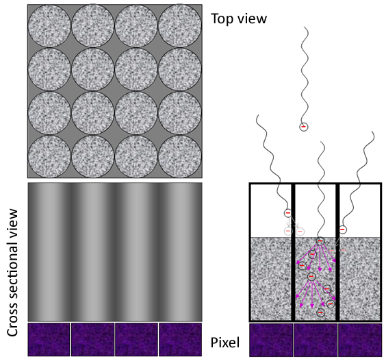
The second approach to guide the light inside the scintallator are silicon mask from semi-conductor industry (e.g. Plasma TVs) filled with a scintillator material:

In the year 2019 the efficiency of these structures is still limited in height and wall-thickness of the "tubes". But there is ongoing work ...

In the year 2019 the efficiency of these structures is still limited in height and wall-thickness of the "tubes". But there is ongoing work ...
Last edited by Klaus on 30.08.2023, 17:06, edited 1 time in total.
Reason: Repaired links to the image
Reason: Repaired links to the image
Re: Structurated Scintillator
30.08.2023, 15:10
Hi Klaus:
Having a deeper understanding of scintillators based on your enlightening post.
Would it be possible for you to share the 5 images mentioned in the preceding post once again?
There might have been an issue with their previous upload.(same issue in Basic principle of the indirect method with Scintillator)
Sincerely appreciate your support.

Best wishs
Colin
Having a deeper understanding of scintillators based on your enlightening post.
Would it be possible for you to share the 5 images mentioned in the preceding post once again?
There might have been an issue with their previous upload.(same issue in Basic principle of the indirect method with Scintillator)
Sincerely appreciate your support.
Best wishs
Colin
Re: Structurated Scintillator
30.08.2023, 17:07
Done - thanks for the advice 
Hosted by iphpbb3.com
Impressum | Datenschutz